Cell Division Mitosis vs Meiosis Explained
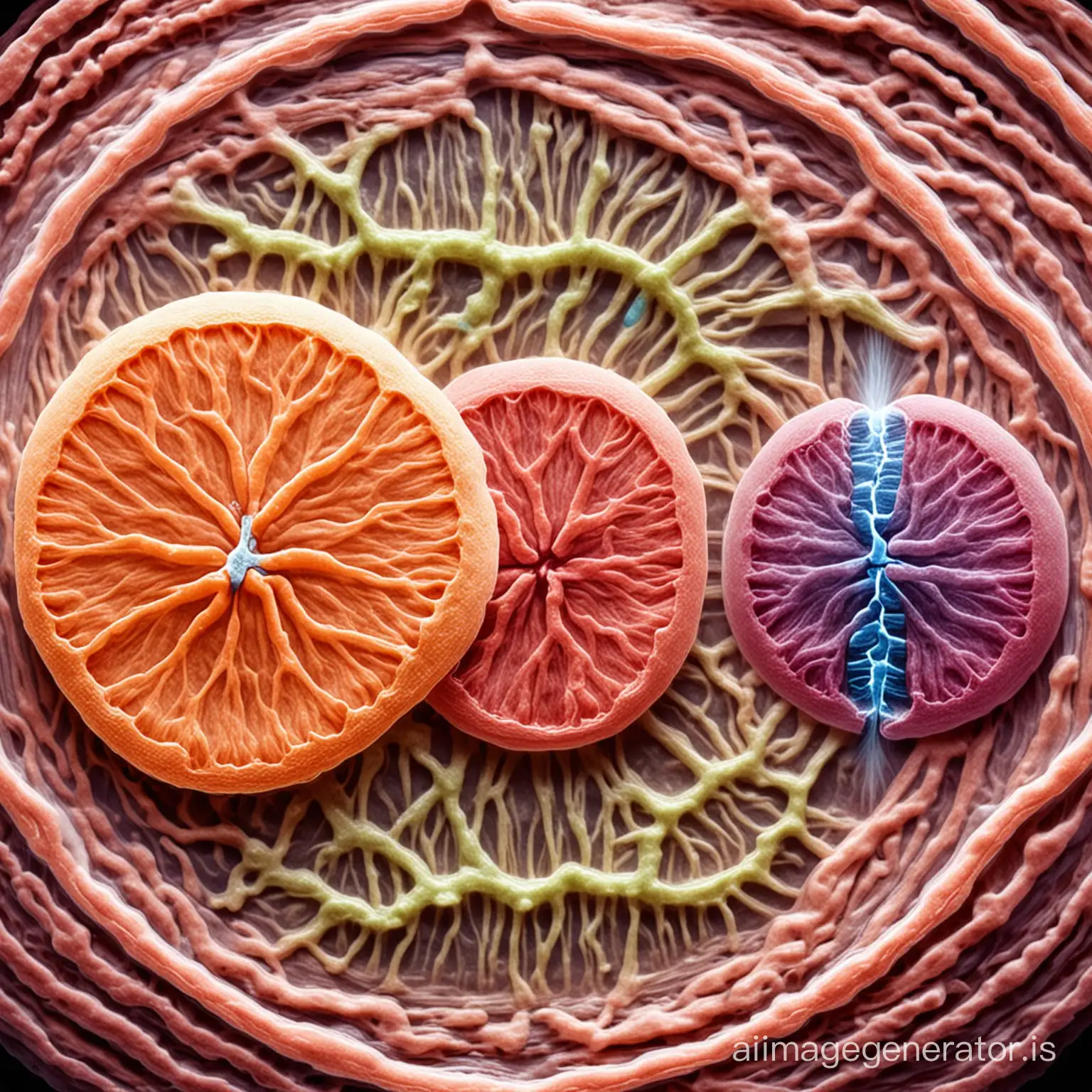
![Mitosis and meiosis division](https://r2.erweima.ai/imgcompressed/compressed_417138b2c967af1ae62de1d54dea32ad.webp)
Image Prompt
Prompt
Mitosis and meiosis division
Model: realistic
Ratio: 1:1
Related AI Images
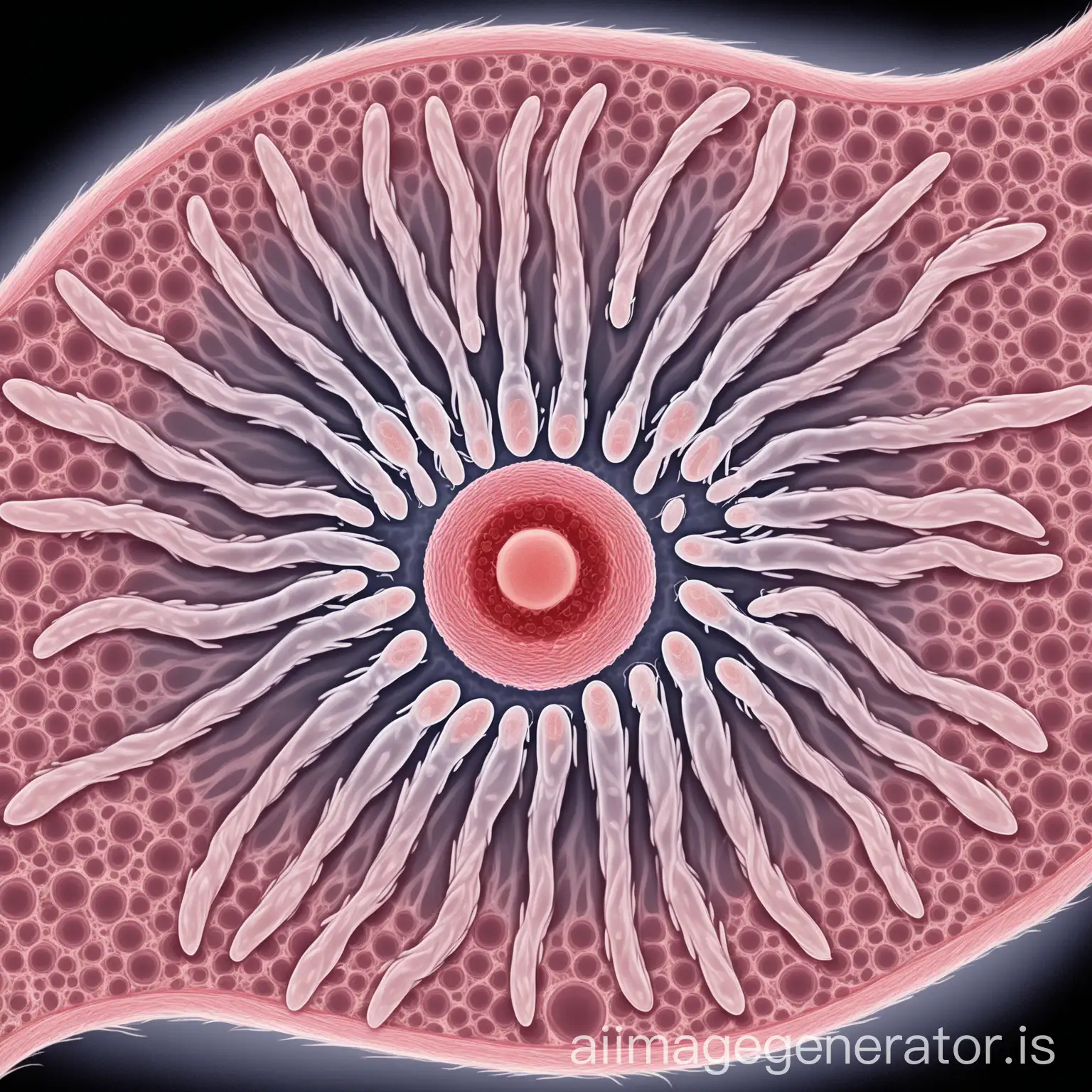
![And after the second division of meiosis, each secondary spermatocyte transformed into spermatids, the precursors of spermatozoa. But that was not all. The spermatids began their last transformation. They prepared for the final battle, developing a tail that would give them the agility necessary to travel through the vast territory of the female reproductive tract. This process is called spermiogenesis.](https://file.aiquickdraw.com/imgcompressed/img/compressed_32915955271c707bf43a29885eb7e1fc.webp)
![plants vs zombies gameplay](https://r2.erweima.ai/imgcompressed/compressed_f8c32536175f5af560e1b7d1815152ce.webp)
![Moloch vs He-Man](https://r2.erweima.ai/imgcompressed/compressed_0a33b2dbb2cb19ebd0dd6299bef02a78.webp)
![MYSQL VS ORACLE DATABASE](https://file.aiquickdraw.com/imgcompressed/img/compressed_a99f9b54175032946925b98967cf5e31.webp)
![Real Madrid vs Barcelona in Santiago Bernabeu Stadium](https://r2.erweima.ai/imgcompressed/compressed_a3bc6eb66093058aa912460acb96c94c.webp)
![generate an cartoon image of a T cell with some random markers on the surface in a use white backgroundnmake the cell bigger and marker smaller put eyes and lips for the cell](https://r2.erweima.ai/imgcompressed/img/compressed_ed5e2de36dcf34be9d8eff7971a59f5e.webp)
![Cancer cell isolated on white background](https://file.aiquickdraw.com/imgcompressed/img/compressed_cab8eb8cc9f701df10b79aaec4ffe191.webp)
![generate an image of a T cell like the red cell in the image with some small random markers on the surface in a white background](https://r2.erweima.ai/imgcompressed/img/compressed_75cac6e3c136e24b0f18c5ada5da1878.webp)
Related Tags
Prompt Analyze
- Subject: Cell Division - Explore the fascinating process of cell division, comparing mitosis and meiosis side by side. Setting/Background: Laboratory Environment - Depict a scientific laboratory setting with microscopes, lab equipment, and cellular models to illustrate the study of cell division. Style/Coloring: Scientific Illustration - Use a detailed and accurate scientific illustration style with vibrant colors to highlight key cellular structures and processes. Action/Items: Chromosomes and Cell Structures - Show the stages of mitosis and meiosis with clear visuals of chromosomes, spindle fibers, and cell membranes undergoing division. Costume/Appearance: Microscopic View - Present a microscopic view of cells undergoing mitosis and meiosis, capturing their intricate structures and dynamic movements. Accessories: Educational Labels - Include educational labels or annotations to explain each stage of cell division, enhancing the image's educational value.